|
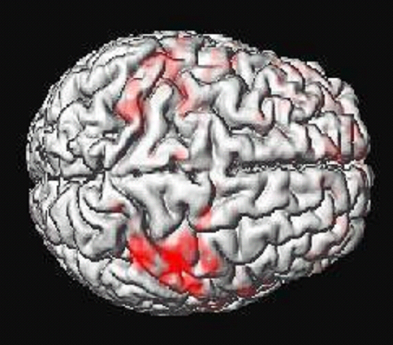
Functional Magnetic Resonance Imaging
(fMRI) allows researchers to obtain images of brain activity
over time, as participants complete various tasks. These images
provide clues about which areas of the brain are related to
cognitive processes, and how different brain regions work together.
The findings from fMRI research shed light on how the brain
is organized.
Available fMRI study and fMRI/TMS study:
Investigation of cerebellar involvement
in cognitive sequencing
Principal Investigator: John E. Desmond, Ph.D., IRB
Protocol number: IRB00328214
Investigation of cerebellar involvement
in alcohol use disorder
Principal Investigator: John E. Desmond, Ph.D., IRB
Protocol number: IRB00337209
What to expect
During the study, an fMRI scanner, like the one in the photo
below will take images of your brain as you complete tasks such
as reading or remembering letters and responding to questions
by a keypad. It is important that you devote all of your effort
to the tasks you complete, in order for the images of your brain
to be meaningful.
fMRI is a very safe, noninvasive imaging
technology. Unlike x-rays, fMRI does not use ionizing radiation.
Instead, fMRI images are generated from a strong magnetic field
and low-power radio-waves, which expose fMRI subjects to much
less energy than x-ray subjects.
You will lie with your back on a table that slides into a horizontal
cylinder in the scanner. Once inside the scanner, it is important
to lie still, as many images of your brain will be taken over
time, and to be useful, the images must line up. In order to
help you stay still and comfortable, pillows will be placed
under and around your head and body in the scanner.
The scanner makes loud beeping noises, so you will be given
earplugs or headphones. You will always be able to communicate
with the researchers, although they will not neccessarily
be able to hear you over the noise made by the scanner. The
researchers will frequently interact with you to make sure that
you are comfortable and to let you know what will happen next.
You should not experience any discomfort. If you do, alert the
researcher and the scan will be stopped if the discomfort is
significant or cannot be alleviated.
|
MRI
scan image
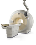
Philips Scanner
|