 |
||
|
Related Sites Kirby
Center for Functional Brain Imaging |
Participate in a NIMLAB TMS Study!
|
We are always
looking for participants for our TMS research studies. Studies require
two 1 hour sessions, one fMRI and one TMS session. In addition to being
paid for their time, participants receive an image of their brain. Transcranial
magnetic stimulation (TMS) is a brain stimulation technique that
involves generating a brief magnetic field in a coil that is placed on
your scalp. The magnetic field passes through the skull and induces a
weak electrical current in the brain that briefly disrupts neural
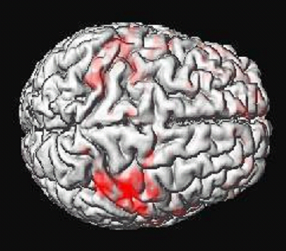
circuits at the stimulation site. Functional Magnetic Resonance Imaging (fMRI) allows researchers to obtain images of brain activity over time, as participants complete various tasks. For the TMS study, we use the results from the fMRI to guide the placement of the TMS coil. |
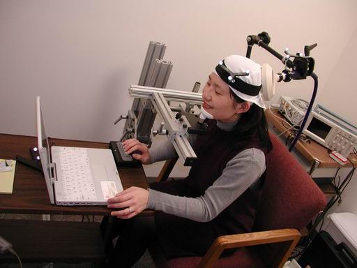
 TMS setup |
|
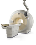
What to expect During the fMRI portion of the study study, an MRI machine, like the one in the photo below, will take images of your brain as you complete tasks such as reading or remembering letters and responding to questions by a keypad. fMRI is a very safe, noninvasive imaging technology. Unlike x-rays, fMRI does not use ionizing radiation. Instead, fMRI images are generated from a strong magnetic field and low-power radio-waves, which expose MRI subjects to much less energy than x-ray subjects. During the TMS session, you will be asked to sit in front of a computer monitor and perform a task for about 20 minutes (with breaks). Periodically while performing the task you will receive a brief magnetic stimulation pulse, and we will record changes in your performance on the task. The magnetic stimulation may cause you to feel a tapping sensation on your head and may cause minor twitching of muscles beneath the coil. If the sensations are too unpleasant for you, you can discontinue the experiment at any time. You will hear a click sound when the magnetic pulse is applied, and you will wear earplugs to reduce the noise. |
|
|
Sign-up! |
 Philips fMRI Scanner |
|
|
|